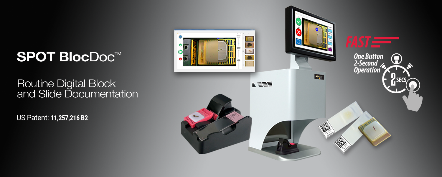
Tissue Pattern Tracking
BlocDoc** provides histology teams streamlined workflows that automatically capture macro images of the cut blocks and raw slides providing Pathologists the images they need to verify their slide examinations.
**- US Patent: US 11,257,216 B2 with International Patents Pending
Does Your Lab Assure Tissue Completeness?
Pathologists are great at making diagnostic determinations on the images and data they view. Now consider for a moment if not all the tissue is being viewed…
… Pathologists cannot diagnose what they cannot see!
Pathology societies around the world have recognized that tissue maybe missing from the final slide or WSI image impacting the diagnosis made 1,2. As a result, accreditation checklists 3,4,5 have been updated to include a requirement for a tissue tracking and review process asking the question...
Does the Lab have a quality check to assure
that relevant tissue is:
1. Being cut from the block
2. Making it onto the raw slide
3. Is present in the final stained slide
4. Is scanned into the whole slide image
Pathologists and pathology directors would like to answer these questions, but in the face of falling reimbursement and rising caseloads, they do not have the resources to manually audit the hundreds of blocks and raw slides that pass through their labs every day.
In the current workflow, what should be done – can't be done.
No Pathologist or laboratory director should be faced with this compromise. Solving this problem isn’t about heroics and staff overtime, it is about having an efficient tool that fits your process and provides clear reference images during the slide examination.
BlocDoc Addresses Pathology Requirements
SPOT Imaging combines 54 years’ experience and a dedicated pathology focus to bring you, BlocDoc™ The New Standard in Paraffin Block and Slide Documentation.
BlocDoc meets accreditation checklist requirements by providing pathologists digital reference images of the cut block and raw slides at the time of slide examination (glass or WSI).
BlocDoc provides enhanced image review of the:
-
-
- Cut Surface of the Paraffin Block
- Below Surface Tissue in the Paraffin Block
- Raw Slide Tissue Levels
-
BlocDoc also provides reference labs an efficient tool for documenting incoming tissue blocks and slides providing an accurate record of what has been received.
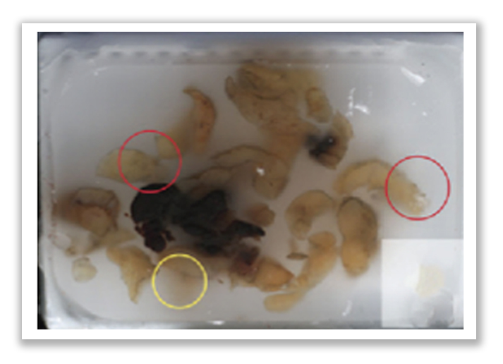
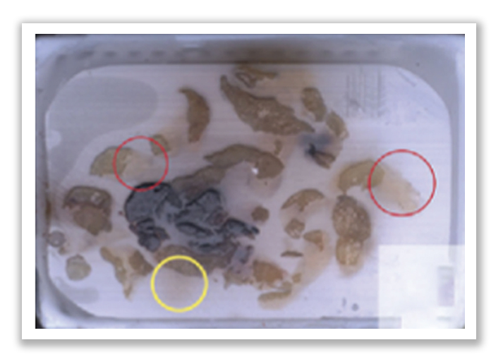
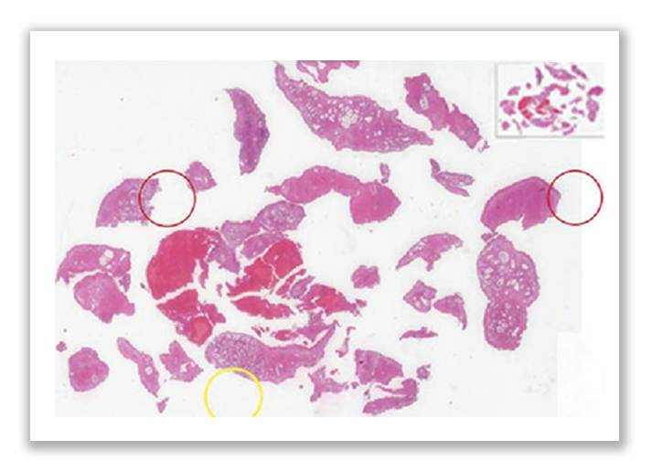
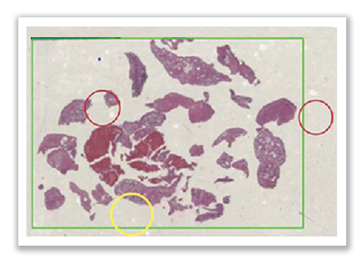
Missing Tissue
Note the subsurface tissue (circled) that is in the paraffin block that did not make it on to the slide for examination. BlocDoc images provide clear digital documentation enhancing both the cut surface and the subsurface tissue.
| BlocDoc image optimized for subsurface tissue. | BlocDoc image optimized for cut surface tissue. |
Areas of missing tissue readily identified In WSI Image |
Areas of missing tissue readily identified In macro image of slide |
1-Filippo Fraggetta,1 Yukako Yagi,2 Marcial Garcia-Rojo,3 Andrew J. Evans,4 J. Mark Tuthill,5 Alexi Baidoshvili,6 Douglas J. Hartman,7 Junya Fukuoka,8 and Liron Pantanowitz7 The Importance of eSlide Macro Images for Primary Diagnosis with Whole Slide Imaging: J Pathol Inform. 2018; 9: 46. Published online 2018 Dec 24. doi: 10.4103/jpi.jpi_70_18
2- Vincenzo L’Imperio, Fabio Gibilisco, Filippo Fraggetta; (2021), What is Essential is (No More) Invisible to the Eyes: The Introduction of BlocDoc in the Digital Pathology Workflow. J Pathol Inform, 12(1), 15. https://www.jpathinformatics.org/text.asp?2021/12/1/32/326167.
3-Liron Pantanowitz, MD; John H. Sinard, MD, PhD; Walter H. Henricks, MD; Lisa A. Fatheree, BS, SCT(ASCP); Alexis B. Carter, MD; Lydia Contis, MD; Bruce A. Beckwith, MD; Andrew J. Evans, MD, PhD; Christopher N. Otis, MD; Avtar Lal, MD, PhD; Anil V. Parwani, MD, PhD Validating Whole Slide Imaging for Diagnostic Purposes in Pathology: Archives of Pathology Laboratory Medicine—Vol 137, December 2013; p.-1710-1722:
4- Peter Hufnagl, Ralf Zwönitzer, Gunter Haroske; Guidelines Digital Pathology for Diagnosis on (and Reports of)Digital Images Version 1.0 Bundesverband deutscher Pathologen e.V. (Federal Association of German Pathologist) diagnostic pathology 2018, 4:266
5- Simon Cross, Peter Furness, Laszlo Igali, David Snead, Darren Treanor Best practice recommendations for implementing digital pathology : The Royal College of Pathologists; Unique document number G162, Date active: January 2018
![]()
![]()
![]()
Budget Positive Implementation…
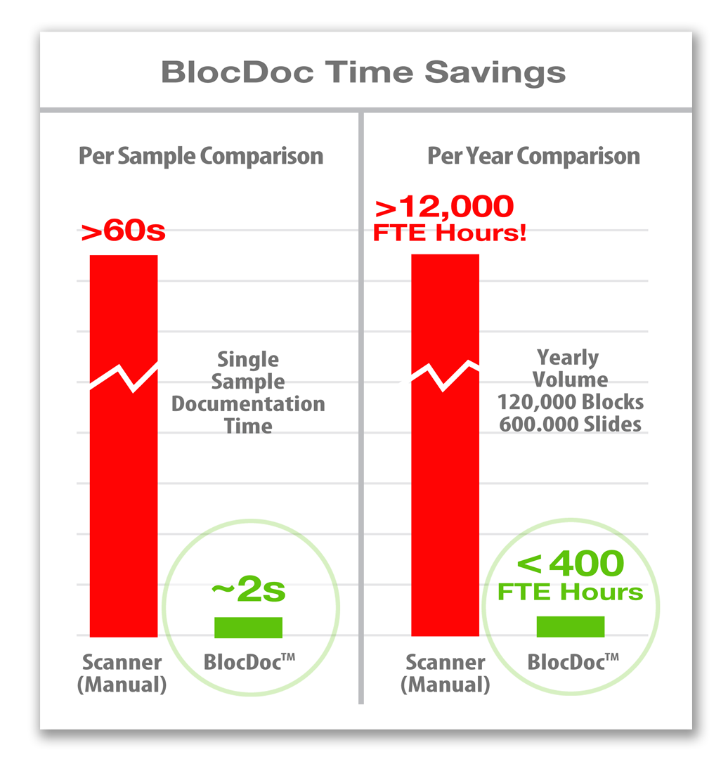
SPOT solutions start with a foundational goal to reduce lab time and pay for themselves in less than a year, making your lab more efficient and allowing your team to stay focused.
Contact SPOT Imaging, get a quote and see how affordable PathSuite can be for your institution.
Streamlined for the Histology Lab
BlocDoc not only meets the needs of the accreditation checklist, it meets the needs of the personnel using it by providing…
 Streamlined Workflow
BlocDoc is an automated image capture system optimized for the Histology workflow. For the microtomist, it is as simple as placing the block onto the sample holder and pushing the trigger button. This is typically a 2 second operation that allows the microtomist to return to their work until they are ready to place the next block or slide.
In the background, BlocDoc takes care of the image acquisition…
• First, a set of reflected and polarized light images are captured
• Next, the sample barcode is captured and decoded
• Lastly, BlocDoc automatically saves the images to the image repository based on the barcode information captured
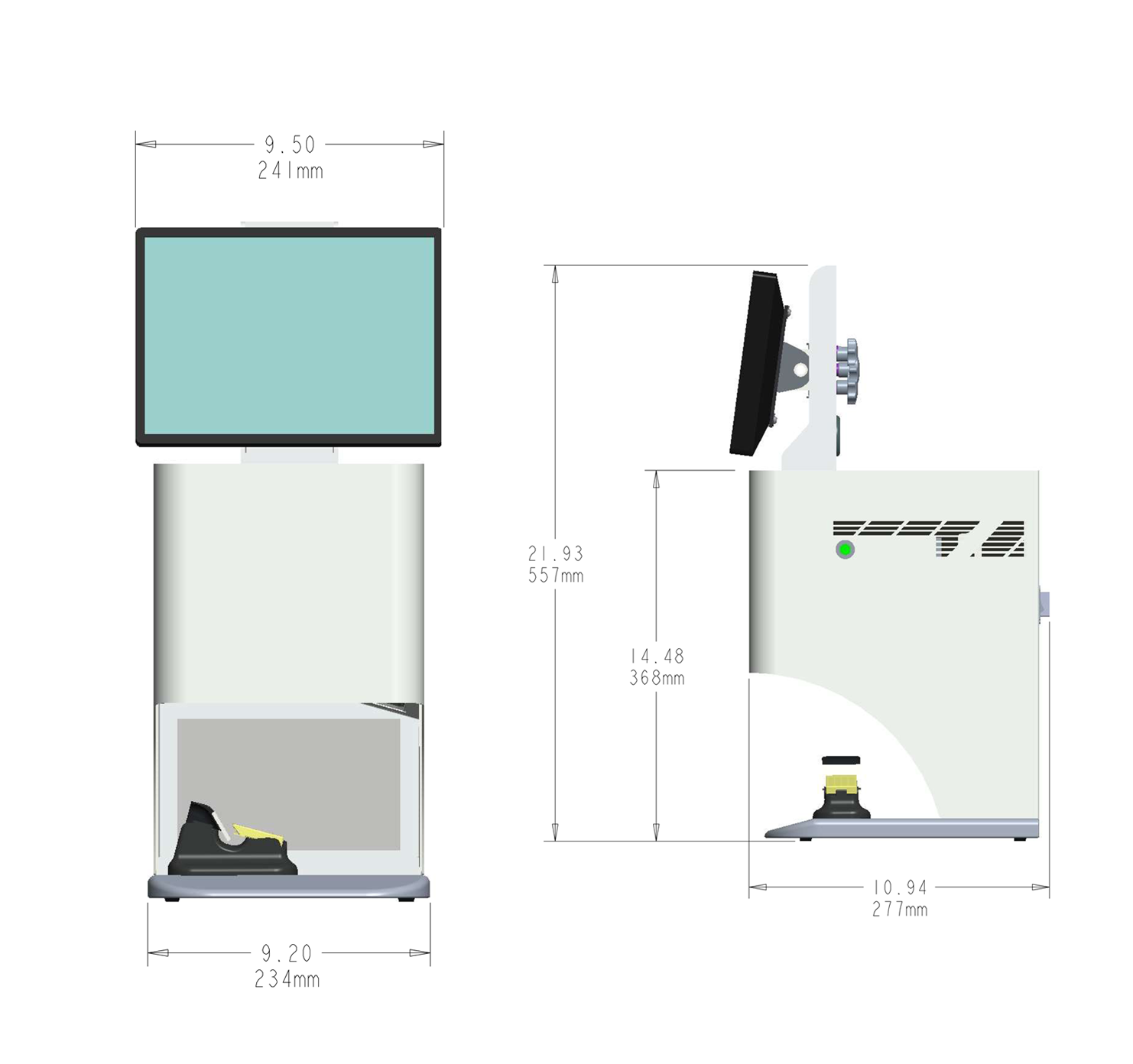
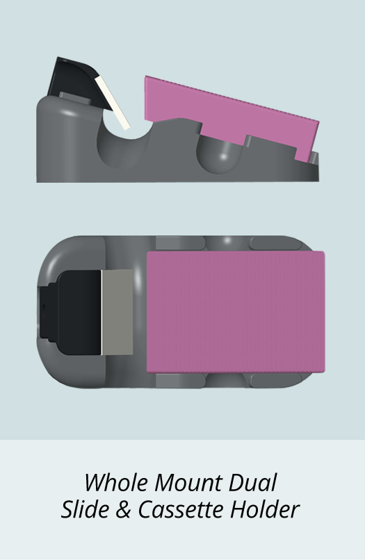
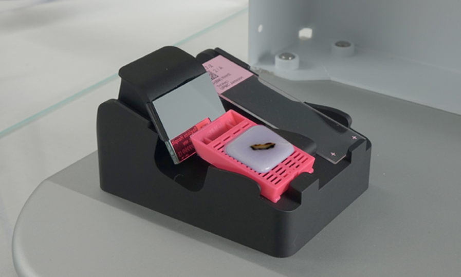
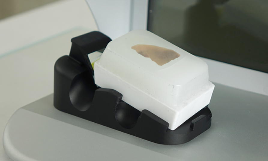
Full Sample Support
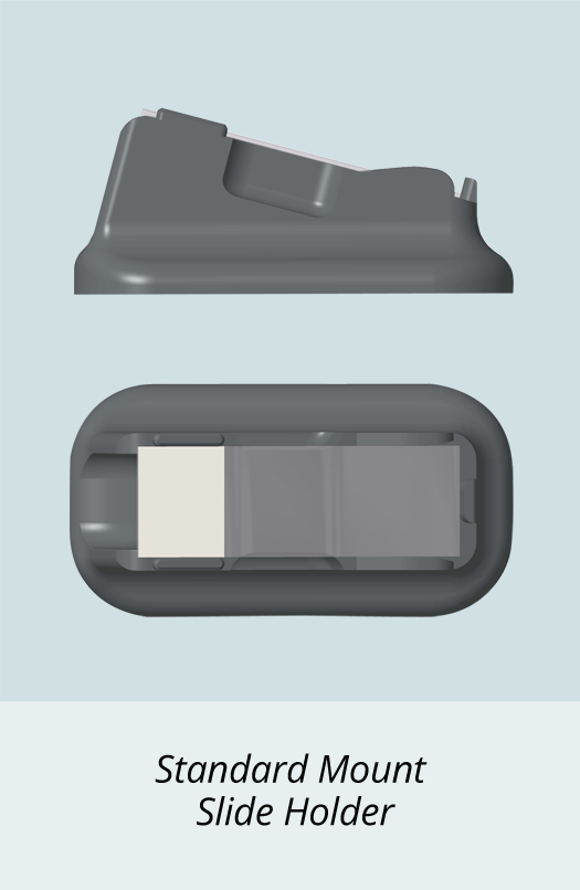
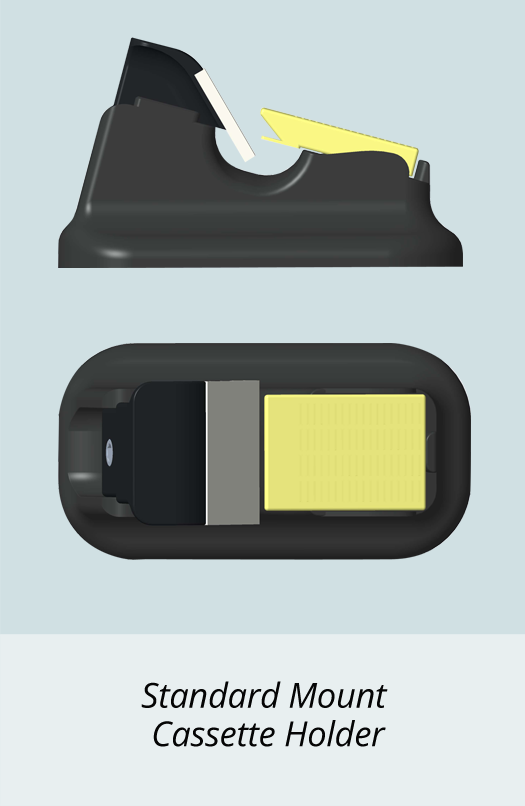
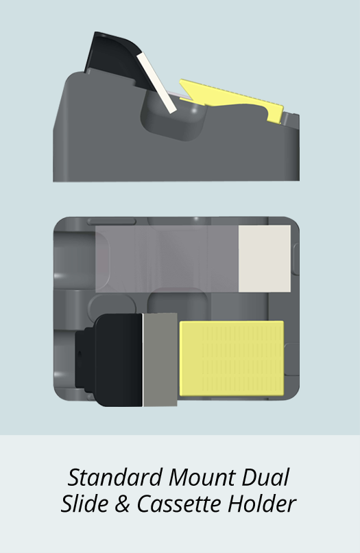
BlocDoc quickly converts standard blocks and slides to one that runs whole mount blocks and slides. The sample holders change out in less than 5 seconds allowing even short runs of whole mounts to be easily accommodated on any BlocDoc instrument.
Showing Cassette & Slide Mounted
Showing Cassette Mounted
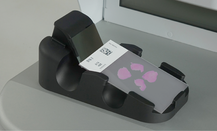
Showing Slide Mounted
Enhanced Images for the Pathologist
BlocDoc’s enhanced images provide the pathologist with a virtual solution that is better than having the physical specimen block in hand.
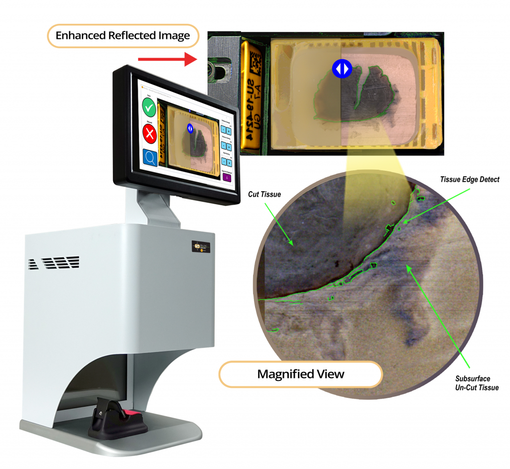
Clarifying the View
BlocDoc’s patented* imaging process, provides enhanced views of the blocks cut tissue. This eases the visualization of what tissue made it to the slide and what tissue is still in the block. The result is greater confidence in calling for recuts or rescans dictated by missing tissue. The BlocDoc system not only makes the tissue review process more convenient it makes the review clear.
*Patent Pending US and Int’l